
Hidden Skin Inflammation: What Your Mirror Can’t Show You (and How to Calm It)

Beauty forms from within. Here’s what’s happening underneath.
Picture a house with a small fire smouldering behind the wall. From the living room, everything looks fine. No flames. No smoke. But if you touch the wall, it is warm.
That’s what hidden skin inflammation can look like. Your mirror says “looks normal.” Our skin scan says something different.
At Skin4Life Beauty Bar in Calgary, I look at skin all day, every day, and here’s what I keep noticing: more and more skin is quietly inflamed. So let’s break it down together. We’ll cover what skin inflammation actually is, why some spots glow red on a scan, what’s causing it, how it holds your skin back, and how to calm it from the inside out.
What is skin inflammation, and why are some spots redder than others?
Inflammation is your body’s security system. When your skin senses trouble (germs, sunburn, rubbing, a harsh product), your immune system sends in the rescue team. To get them there fast, the tiny blood vessels near the surface open wider, kind of like widening a road so more ambulances can get through. That brings extra blood, immune cells and nutrients to the spot. More blood close to the surface also means skin that looks and feels redder and warmer. The medical word for that redness is erythema.
A short burst of inflammation is a good thing. In a normal wound, it calms down on its own within one to two weeks [2]. The trouble starts when the alarm just keeps ringing.
So why do some parts of the face look redder?
- Some faces are just built that way. Cheeks and nose have more blood vessels close to the surface and thinner skin, so colour shows through more.
- The spots that get the most attention. Areas you rub, that get the most sun, or that take the biggest hit from acids, scrubs and strong products usually react first.
- Breakouts and touchy patches. Active or recent breakouts and reactive areas draw extra blood flow while the skin repairs itself.
- Heat and flushing. Hot showers, stress and spicy food can open up those vessels for a while.
So what is the redness telling you?
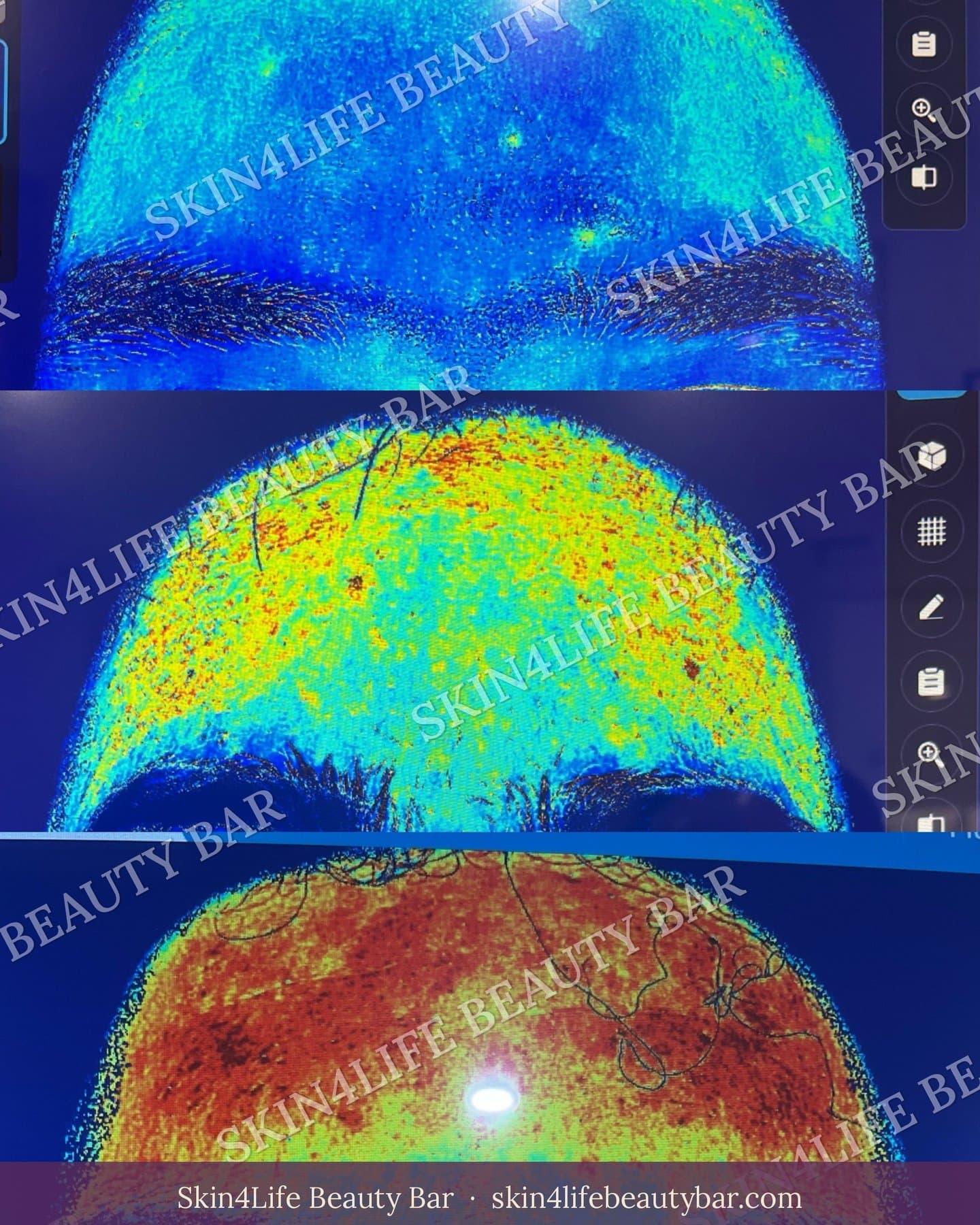
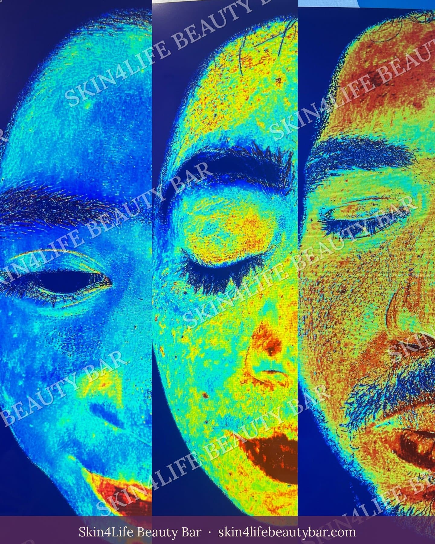
A warm colour on the scan is basically your skin saying, “the alarm is on in this spot.” Your barrier may be stressed, and your skin is more reactive than it should be. In my experience, lots of these areas look only slightly pink, or totally normal, in the mirror. That’s why I call it hidden inflammation: the scan sees it first.
A quick honest note: a skin scan shows a redness signal. It’s a really helpful clue, but it’s not a medical diagnosis. I always read it together with your skin history, how your skin feels, and your lifestyle.
Short-term vs. long-term: when the alarm never switches off
Scientists call this slow-burn, long-term inflammation inflammaging (inflammation plus ageing). Researcher Claudio Franceschi introduced the term in 2000. Regular, short-term inflammation fights off injury or infection. Inflammaging doesn’t switch off, and it wears things down over time [1]. In skin it’s often silent: no pain, no swelling, just a bit pink, a bit sensitive, a bit dull, and slow to heal.
What I see in the treatment room every day
More and more clients walk in with skin that’s working overtime. When I look closely, the same four things keep showing up:
1. Too many exfoliating acids. A scrub on Monday, glycolic pads on Tuesday, a BHA serum on Wednesday, all “for glow.”
2. Strong actives stacked on top of each other. Retinol, vitamin C, acids and peels in the same routine, often with no rest day.
3. Late nights. Your skin does a lot of its repair work while you sleep.
4. A diet that doesn’t help calm skin. Lots of sugar, fried and ultra-processed food, and not enough colourful plants and healthy fats.
Here’s an everyday example. Think about washing your hands thirty times a day with strong soap. They’d end up red, tight and sore. Your face reacts the same way to daily over-exfoliating. The skin barrier (the outer layer that keeps water in and irritants out) gets damaged, water escapes, irritants sneak in, and your immune system keeps jumping into action. Skin doctors link a damaged barrier with ongoing inflammation [12].
These are my own observations from the treatment room, not a study. But the science backs up each one, so let’s go through it.
What keeps skin inflamed? Stress from the inside and the outside
From the inside
- Sleep. In a study of 60 healthy women, the poor sleepers had a weaker skin barrier. After researchers stripped the top layer of skin with tape, the good sleepers rebuilt about 30% more of their barrier within 72 hours, and they bounced back from UV-caused redness better too [5]. It was a small study, so it shows a link, not proof.
“Chronic poor sleep quality is associated with increased signs of intrinsic ageing.”
— Oyetakin-White and colleagues, dermatology researchers, Clinical and Experimental Dermatology, 2015 [5]
- Stress. Researchers talk about a “brain–skin connection”: stress signals can switch on inflammation in your skin and speed up ageing [6]. Ever had a breakout right before a big event? That’s the connection at work.
- Food. Lots of sugar, plus grilled or fried food, is linked with a build-up of advanced glycation end products, or AGEs. These form when sugar sticks to proteins like collagen. Think of toast or caramel browning, except it’s happening slowly inside your skin and making it stiffer and worse at repairing itself [7]. On the bright side, healthy fats like omega-3 may help support your barrier and calm inflammation, although most of that evidence comes from lab and animal studies so far [7].
From the outside
- Sun (UV). UV light creates oxidative stress (more on that in a second) and triggers inflammation [8].
- Pollution. Tiny particles, ozone and smoke can create free radicals, trigger inflammation and weaken your barrier. In one group of older women, more exposure to particle pollution was linked with more pigment spots on the face and deeper lines from nose to mouth (the nasolabial folds) [8].
- Dry, cold air and indoor heating. Calgary winters pull moisture out of your barrier. That’s tough on healthy skin and even tougher on skin that’s already inflamed.
- Over-treating. As we saw above, too many acids, scrubs and strong products can be the biggest stress of all.
Notice something? A lot of these triggers share the same troublemaker: free radicals.
Free radicals: the spark behind the fire
Think of a free radical as an unstable oxygen molecule that’s missing an electron and desperate to get one. It steals from the nearest healthy molecule (like collagen, skin fats or DNA), and then that molecule becomes unstable too. It’s a chain reaction, the same thing that turns a cut apple brown or makes iron rust.
Your body makes some free radicals naturally and keeps antioxidants on hand to neutralise them. The trouble starts when too many show up: UV, pollution, smoke, stress, poor sleep or a sugar overload. That tipping point is called oxidative stress. One review calls it the leading cause of photoaging (sun-related skin ageing) [8].
Here’s how it links to inflammation. Free radicals and inflammatory signals switch on enzymes called matrix metalloproteinases, or MMPs. Think of them as tiny scissors that cut up collagen and elastin, the proteins that keep skin firm and bouncy. The broken pieces fuel more inflammation, which makes more free radicals [2,8]. Researchers call this a self-perpetuating cycle. The two keep each other going, like two people fanning the same flame.
How hidden inflammation stops your skin from getting stronger
Inflamed skin is busy putting out fires, so it has no energy left to build anything new. Here’s what that costs you:
1. Slower recovery. Ongoing inflammation stalls repair. Too many inflammatory signals and free radicals damage the skin’s support structure and slow down new collagen [2].
2. More dark marks, especially in deeper skin tones. Inflammation turns up your pigment cells (melanocytes), so they make more melanin and pass it to nearby skin cells. The result is post-inflammatory hyperpigmentation, or PIH: the dark marks left behind after skin has been irritated or inflamed [3].
“Post-inflammatory hyperpigmentation ‘tends to affect darker skinned patients with greater frequency and severity.’”
— Erica Davis, MD, and Valerie Callender, MD, Journal of Clinical and Aesthetic Dermatology, 2010 [3]
The same review explains that pigment sitting in the upper layer can take months to years to fade, while pigment that drops deeper looks blue-grey and may be permanent [3]. Melasma is more complicated, since hormones, UV and genetics are all involved. But dermatologists have also noticed that melasma skin has more, and larger, blood vessels than the skin around it [4]. Red and brown often travel together.
3. Loss of firmness and bounce. Remember those MMP scissors? Inflammation and free radicals turn them up and slow new collagen, so skin slowly loses its elasticity [2,8].
4. Higher risk with stronger treatments. Cosmetic procedures are a known trigger for PIH, and irritation can make it worse [3]. Even microneedling leaves tiny channels that can make skin more prone to irritation, infection and PIH [13]. Treating skin that’s already inflamed is like starting a race with a fever.
5. Lasting sensitivity. Once your barrier stays weak, the alarm goes off more easily. Products that used to feel fine start to sting [12].
6. Less ability to fight germs. In inflamed eczema skin, researchers found much lower levels of two of the skin’s own germ-fighting proteins (LL-37 and human beta-defensin-2). That may be why inflamed, eczema-prone skin picks up Staphylococcus aureus (staph) infections more easily [9].
7. Faster ageing. Long-term inflammation is one of the main drivers of ageing skin, the inflammaging we talked about earlier [1].
Good inflammation vs. silent inflammation: why it decides your results
Here’s something that surprises many clients: in a clinic, we sometimes cause inflammation on purpose.
Treatments like microneedling, peels and lasers work through controlled injury. They create tiny, planned wounds, and your skin reads them as a signal to repair itself. Immune cells release growth factors (little messengers that tell skin to rebuild), fibroblasts (your collagen-making cells) get to work, and the new tissue gradually remodels [13]. Here, inflammation is the starting gun for renewal. It’s planned, measured, done on prepared skin, and followed by recovery.
Think of a workout. Training creates tiny, controlled damage in your muscles, and with rest and good food you come back stronger. Train every single day with no rest and you get injured. Chronic inflammation is overtraining for your skin. It’s unplanned, it never stops, and your skin never gets the recovery phase.
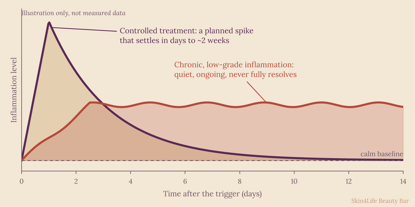
This is why chronic inflammation gets in the way of good results:
- Your skin’s repair crew is already busy, so a treatment piles a second spike on top of the first instead of starting from calm.
- Redness, dark marks and downtime become more likely, especially in deeper skin tones.
- Results are weaker, because new collagen is being built in an environment that’s breaking collagen down.
That’s why I sometimes tell clients the best treatment this month is no treatment: calm the skin first, get a stronger result later. A scan like the one below helps guide that conversation.
How to calm skin inflammation from the inside out
Start with the inside
- Protect your sleep. Aim for a steady 7 to 9 hours. That’s the range the good sleepers in the study above fell into [5].
- Turn your stress down a notch. A slow walk, deep breathing or a quiet evening ritual takes pressure off your skin as well as your mind [6].
- Eat to calm. Build meals around colourful vegetables and fruit, and add omega-3 foods like salmon, walnuts and flaxseed. Go easier on sugar and on fried or grilled food [7].
- Wear sunscreen every day. It stops the UV spark before it starts [8].
Then choose calming ingredients
Here are the ingredients I like for inflamed or reactive skin, and what the research actually says:
| Ingredient | What it does, in plain words | What the evidence says |
|---|---|---|
| Niacinamide (vitamin B3) | Helps skin build its own barrier fats, calms inflammatory signals and slows pigment from moving up to the skin’s surface. | Well tolerated. In a split-face trial, 5% twice daily for 8 weeks reduced hyperpigmentation, while 2% did not reach significance [10]. |
| Centella asiatica (madecassoside, asiaticoside) | A traditional soothing plant. Its active compounds (madecassoside and asiaticoside) are best known for calming and repairing skin. | Best known for treating skin conditions, but much of the research is lab-based or early-stage [11]. |
| Ectoin | A protective molecule from organisms that survive harsh environments. It holds a thin layer of water around skin proteins and cell membranes, which helps hydration and barrier strength. | A review of 6 clinical studies found ectoin creams (5.5 to 7%) were well tolerated, with less itch and dryness in atopic dermatitis. Certainty was low to moderate, and the review was funded by an ectoin manufacturer [12]. |
| Antioxidants (vitamins C and E) with daily SPF | Antioxidants neutralise free radicals, while SPF blocks the UV that creates them. | Researchers suggest UVA-protective sunscreens plus antioxidants may help against pollution and UV stress [7,8]. |
| Barrier fats (ceramides, cholesterol, fatty acids) | Refill the “mortar” between skin cells so water stays in and irritants stay out. | The standard approach to barrier repair. Niacinamide also boosts the skin’s own production of these fats [10]. |
A simple reset you can start tonight
1. Pause the extras. Take a break from scrubs, stacked acids and high-strength products while your skin is reactive.
2. Cleanse gently with a mild, soap-free cleanser and lukewarm (not hot) water.
3. Support your barrier with a moisturiser that contains barrier fats, and add niacinamide, centella or ectoin.
4. Protect daily with SPF, and keep your routine simple for 2 to 4 weeks.
5. Bring things back slowly. Add one active at a time, ideally with a professional’s guidance.
Conclusion: calm first, then strengthen
Healthy skin isn’t about doing more. It’s about knowing when to stop, protecting your barrier, and giving your body what it needs to repair. Inflammation isn’t the enemy. Planned, it can renew your skin. Silent and constant, it quietly holds your skin back and limits what any treatment can do for you.
If your skin looks fine in the mirror but feels sensitive, dull or slow to heal, there could be more going on underneath.
Curious what’s happening underneath your skin? Book a facial at skin4lifebeautybar.com and get a skin scan to see what’s going on below the surface.
This article is for education and does not replace advice from your doctor or dermatologist. If you have a persistent rash, pain, swelling or signs of infection, please see a medical professional.
Book your consultation
Book at Skin4LifeCurious about skincare science, at-home and in-clinic treatments, and decoding the latest trends? Follow along on Instagram @skin4life_beautybar.
References & Citations
- Haque A, Woolery-Lloyd H. Inflammaging in dermatology: a new frontier for research. J Drugs Dermatol. 2021;20(2):144-149.
- Zhao R, Liang H, Clarke E, Jackson C, Xue M. Inflammation in chronic wounds. Int J Mol Sci. 2016;17(12):2085. https://www.mdpi.com/1422-0067/17/12/2085
- Davis EC, Callender VD. Postinflammatory hyperpigmentation: a review of the epidemiology, clinical features, and treatment options in skin of color. J Clin Aesthet Dermatol. 2010;3(7):20-31.
- Talakoub L, Wesley NO. The hidden vascular component of melasma. MDedge Dermatology, Aesthetic Dermatology Update (clinician commentary summarising published studies). https://mdedge.com/content/hidden-vascular-component-melasma
- Oyetakin-White P, Suggs A, Koo B, et al. Does poor sleep quality affect skin ageing? Clin Exp Dermatol. 2015;40(1):17-22.
- Chen Y, Lyga J. Brain-skin connection: stress, inflammation and skin aging. Inflamm Allergy Drug Targets. 2014;13(3):177-190.
- Cao C, Xiao Z, Wu Y, Ge C. Diet and skin aging: from the perspective of food nutrition. Nutrients. 2020;12(3):870. https://www.mdpi.com/2072-6643/12/3/870
- Parrado C, Mercado-Saenz S, Perez-Davo A, Gilaberte Y, Gonzalez S, Juarranz A. Environmental stressors on skin aging: mechanistic insights. Front Pharmacol. 2019;10:759.
- Ong PY, Ohtake T, Brandt C, et al. Endogenous antimicrobial peptides and skin infections in atopic dermatitis. N Engl J Med. 2002;347(15):1151-1160.
- Boo YC. Mechanistic basis and clinical evidence for the applications of nicotinamide (niacinamide) to control skin aging and pigmentation. Antioxidants. 2021;10(8):1315. https://www.mdpi.com/2076-3921/10/8/1315
- Tan SC, Bhattamisra SK, Chellappan DK, Candasamy M. Actions and therapeutic potential of madecassoside and other major constituents of Centella asiatica: a review. Appl Sci. 2021;11(18):8475. doi:10.3390/app11188475
- Kauth M, Trusova OV. Topical ectoine application in children and adults to treat inflammatory diseases associated with an impaired skin barrier: a systematic review. Dermatol Ther (Heidelb). 2022;12:295-313. https://link.springer.com/10.1007/s13555-021-00676-9
- Tehrani L, Tashjian M, Mayrovitz HN. Physiological mechanisms and therapeutic applications of microneedling: a narrative review. Cureus. 2025;17(3):e80510. doi:10.7759/cureus.80510
This content is for educational purposes and does not constitute medical advice.